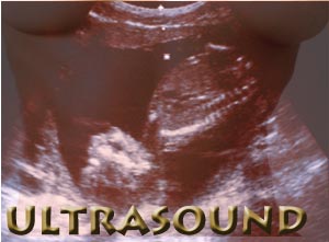
Pregnancy Ultrasound

It's an exciting day for an expectant mother when she can get a glimpse of the little baby growing within her. She can watch the baby's profile as well as the beating of the tiny heart. Ultrasonography has become a very useful diagnostic tool in obstetrics. During pregnancy, fetal development and female pelvic organs can be observed with an ultrasound device. Fetal heart movements can be assessed and measurements can be made accurately with the help of the pregnancy ultrasound. Such measurements are vital in assessing the gestational age, size and growth of the fetus.
Pregnancy ultrasound
High frequency sound waves are transmitted from the ultrasound machine. An image is formed when these sound waves rebound from the body structure. The image is itself an echo of the sound wave transmitted by the ultrasound machine. The echo that bounces back helps to identify where the object is located, how big it is, it's shape and whether it's internal consistency is liquid, solid or both. The echo takes longer to return if the object is far away and is heard sooner if the object is close by.
A picture is reproduced on the computer screen, converting the sound waves by measuring them at once. As the ultrasound machine senses the echo, it places a dot on the monitor. The position of the dot depends on the return echo. A strong echo from deep within the womb will be whiter and gray if the echo is nearer the top of the monitor screen. Ultrasonography machine consists of:
- The transducer
- The monitor
- The machine on the whole
Transducer: Transducer is held in the hands of the person performing the ultrasonography examination. A crystal from within it emits ultrasound beams and waits to listen for echoes. The transducer is moved on the area that needs to be examined.
Monitor: The TV screen that displays the moving images of the fetus.
The machine: The Ultrasonography machine transfers echoes from echoes into images that are seen on the monitor.
Pregnancy ultrasound system
Standard ultrasound is routinely conducted on most pregnant women to evaluate the growth and development of the baby as well as identify a multiple pregnancy. Advanced ultrasound is useful in tracing any suspected problem.
Pregnancy ultrasound with 3-D imaging provides more detailed images and throws up more information on birth defects. 3-D Ultrasound machines use specially designed probes and software to generate 3-D images of the growing baby. Doppler ultrasound measures waves as they bounce off moving objects. Fetal echocardiography aids in detecting any possible congenital heart defects in the fetus.
Trans abdominal pregnancy ultrasound: Trans abdominal ultrasound is the most commonly- used method of pregnancy ultrasonography. A conducting gel is applied on the abdomen as it aids transmission of the sound beam from the transducer to the skin. The transducer is moved on the abdomen.
Trans vaginal ultrasound: Your bladder needs to be empty to get this scan done. An ultrasound probe is placed in the vagina and the scan is performed. The structures can be visualized clearly and precisely when compared to a trans abdominal scan. The only shortcoming is ultrasound beam from the probe does not travel as far into the body when compared to the trans abdominal type.
Preparing for pregnancy ultrasound
If you have chosen to go in for trans-abdominal ultrasound, then you may have to drink plenty of water. An hour before the test you will be asked to drink as much as 3 -4 glasses of liquid. This happens in the case of ultrasonography in the first trimester. A full bladder eliminates pockets of air between your uterus and bladder and ensures clear images. As the pregnancy advances, the amniotic fluid offers the medium and helps push the bladder down. This allows the uterus stretch out directly against the abdomen.
You need not remove your dress but will have to expose your abdomen alone. The procedure can take about 15 to 45 minutes. Remember not to urinate till the pregnancy ultrasound is through. Hold your bladder, even if you have the urge to urinate. There may be some discomfort from pressure on the full bladder. The ultrasound waves do not create any sensations.
Uses of pregnancy ultrasound
Your physician will decide on the frequency of the ultrasound to be performed during the course of your pregnancy. In general a scan during the pregnancy is recommended to substantiate a normal pregnancy.
- Gestational sac can be seen as early as four and a half weeks and the yolk sac at about five weeks. The embryo can be measured at about five and a half weeks and the ultrasound can confirm the position of the pregnancy during this time.
- Heartbeat of the fetus can be detected by seven weeks and missed abortions can be ruled out at this stage if you have spotting.
- Molar pregnancies and ectopic pregnancies can be ruled out using ultrasound if there is vaginal bleeding at this stage.
- Abdominal circumference can be measured with pregnancy ultrasound. This important measurement in late pregnancies as this reflects on the fetal weight and size more than the age.
- Any type of fetal malformation such as chromosomal disorders and spina bifida can be diagnosed when the fetus is 20weeks of gestational age.
- Pregnancy ultrasound is vital in locating the placenta and finding its lower edges.
- Pregnancy ultrasound aids in assessing the amount of amniotic fluid.
- Vital measurements such as femur length and biparietal diameter provide information on the gestational age.
Benefits of pregnancy ultrasound
Pregnancy ultrasound is non-invasive and does not entail exposure to x-ray radiation. The results are produced quickly after the test is done and the test itself is not too long. The ultrasonography machine creates a moving image. This makes it easy for the doctor to follow the fetal movements. But pregnancy ultrasounds must be undertaken only on the advice of a health worker or doctor. It is not advisable to expose a person to unnecessary ultrasound procedures.
Top of the Page: Pregnancy Ultrasound
Tags:#pregnancy ultrasound #Ultrasonography
 Planning Pregnancy
 Pregnancy Diet and Exercise
Healthy Weight Gain in Pregnancy
Pregnancy Food Cravings
Pregnancy Heartburn
Improving Fertility after 35
Ovulation Test Strips
Pregnancy Symptoms
Early pregnancy cramping
Early pregnancy Test
High Risk Pregnancy
More on Pregnancy
 Natural Childbirth
 Premature Birth
Pregnancy Massage
Pregnancy Problem
Pregnancy Due Date Calculator
Amniocentesis
Twin Pregnancy
Ectopic Pregnancy
Pregnancy Morning Sickness
Molar Pregnancy
Pregnancy Ultrasound
Constipation during pregnancy
Insomnia during Pregnancy
Smoking during Pregnancy
Teen Pregnancy
Exercise during pregnancy
Late Term Abortion
Sign of Miscarriage
Pregnancy Fatigue
Pregnancy Gas
Pregnancy sciatica
Belly Wrap after Pregnancy
Emotional Health during Pregnancy
Lactating Mother
Infertility
Recurrent Miscarriage
Pregnancy Varicose Veins
Postpartum Depression
Epidural
Premature Infant
Top of the Page: Pregnancy Ultrasound
Popularity Index: 100,814

