Diagnostic mammogram

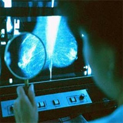
Mammography is an imaging process that uses low-dose x-rays to examine the breasts. A mammogram image or picture can help in early diagnosis of cancer. Mammograms are essential for women who have had breast cancer or those with a genetic history of breast cancer.
According to the guidelines laid by the U.S. Department of Health and Human Services (HHS), the American Cancer Society (ACS), the American Medical Association (AMA) and the American College of Radiology (ACR), annual diagnostic mammograms are recommended for women above age 40. Mammogram screening aids in diagnosing breast diseases or lumps.
Mammogram procedure
Mammogram images are taken with a mammography unit, which houses the tube that produces the x-rays. This equipment is specifically engineered to x-ray the breast. It is equipped with equipment that holds and compresses the breast and positions it so that mammogram images are obtained at different angles.
Mammogram procedure requires the breast to be compressed because it evens out the breast thickness so that all of the tissue can be imaged. In this way, small abnormalities are not hidden by overlying breast tissue. Compression of the breast for mammogram screening also reduces x-ray scatter so as to obtain a sharp picture.
It is essential to position the breast correctly during mammogram screening otherwise the entire breast will not be imaged. The patient must be informed of all the steps of the mammography process. You will be asked to position your breast on a small flat plate with an x-ray plate under it. Another flat plate is positioned above which is used to compress the breast.
The patient is required to remain absolutely still and hold the breath to eliminate image blurring during the mammogram procedure. The technologist goes behind the glass shield and asks you to change positions slightly between mammogram images.
Mammogram Screening
Mammogram screening and consequent images allow a physician to detect small tumors. Screening mammography helps detect small abnormal tissue growths that are confined to the milk ducts. This is Ductal carcinoma in situ (DCIS) and is generally detected with a diagnostic mammogram. An effective radiation dose used for mammogram screening is about 0.7 mSv.
Sometimes, abnormal mammogram pictures are seen and these cases are followed up with other tests. Women must indicate to the technologist in case they are pregnant before undergoing a mammogram screening. A patient should inform the technologist is there is any significant pain during the mammogram procedure.
Digital Mammogram
The digital mammogram utilizes a digital receptor and computer to produce detailed images of the breast. This electronic manipulation of mammogram images allows for fast stereotactic biopsy. The physician can magnify or zoom in to different parts of the breast tissue without taking any additional mammogram images.
Digital mammograms display the images immediately on the monitor and save considerable time and effort. Digital mammograms show improved contrast between dense and other breast tissue. You can rectify under or exposed films without repeating the mammogram screening procedure.
In conventional mammograms, the images are recorded on film and viewed through a light box. Digital mammogram images are stored as digital pictures which can be reviewed on the monitor. These images can be transmitted easily for remote consultation. The magnification, brightness, contrast and orientation can be altered to view the breast tissue clearly. Digital mammogram pictures are more expensive as compared to standard film mammogram images.
Mammogram result
A study of mammogram results has indicated that obese women are at increased risk for having abnormal mammogram results erroneously. This 'false positive' mammogram result may be attributed to poor mammogram image clarity on account of thicker volume of breast tissue. Since the breast size, shape and tissue density differs for each woman and therefore the technologist must pay attention to the mammogram image carefully.
To get an optimal mammogram image, larger breasts may require higher x-ray doses. A good mammogram image must indicate sufficient contrast to enable detection of lumps, cysts and other tissue masses.
Abnormal mammogram
Mammogram results are categorized into 5 types by the American College of Radiology. A '0' means that the mammogram screening needs to be repeated. A '1' indicates a negative mammogram result. A '2' indicates identification of an abnormality that may or may not be malignant. A '3' indicates an abnormality that is most likely to be benign.
A '4' is indicative of a suspicion of cancer whereas a '5' is a high chance of cancer. Abnormal mammograms can be the resultant of solid-dense areas or calcifications. They are not indicative of cancer though.
Calcification mammogram
Calcification can be easily detected on the mammogram picture. We carry a detailed article on Breast Calcification that will help you gain some insight into this condition. Calcification mammograms that indicate tiny, irregular deposits can be a cause for worry.
Top of the Page: Diagnostic mammogram
Tags:#mammogram #diagnostic mammogram #mammogram result #free mammogram #abnormal mammogram #mammogram picture #mammogram screening #breast cancer mammogram #mammogram image #digital mammogram #calcification mammogram #mammogram procedure

Cancer Staging and Grading
Mammogram - Breast Xray
Breast Ultrasound
Breast MRI
Breast Augmentation
More on Breast cancer

Stress and Breast Cancer
Breast Density and Breast Cancer Risk
Lower your Breast Cancer Risk
Breast Cancer Myths
Mastitis
Breast Cancer Awareness
Preventing Breast Cancer
Breast Self Exam
Breast Cancer Chemotherapy Treatment
Palpable Breast Mass
Breast Cyst
Breast Cancer Symptoms
Breast Calcification
Breast Cancer Treatment
Mastectomy
Top of the Page: Diagnostic mammogram
Popularity Index: 101,412